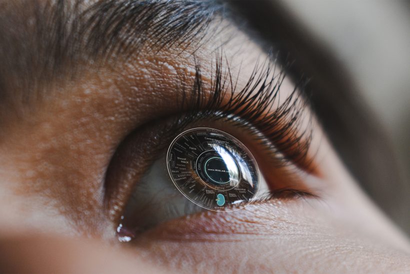
In this course, students will learn through studying extensive iridology research. Iridology itself is the study of the iris of the eye, specifically the colored portion. Even in the modern era, many health practitioners still make use of iridology to help better diagnose patients.
These are the specifications of Iridology Specialization Course:
| 1. Awarding Institution / Body: | |
| 2. Teaching Institution: | Online and distance learning, with tutor support |
| 3. Programme Accredited by: | |
| 4. Final Award: | Compulsory course for the Bachelors in Naturopathy and Holistic Medicine degree |
| 5. Programme title: | Iridology |
| 6. Course Code and level: | BNHM6022 – Bachelor Level |
| 7. Duration of programme: | One trimester or 12 weeks |
| 8. Total number of study hours: | Equivalent to 6 US credits or 60 clock hours of study |
| 9. Enrollment requirements: | Students enrolled in the Bachelor’s degree |
| 10. Enrollment date: | Third-year, Second trimester of Bachelor’s degree |
| 11. Fees: |
Lessons
- Lesson One – Introduction to Iridology
- This first level introduces the roots of Iridology going back to findings in Tutankhamen’s tomb toward current technological advancements and developments. Results obtained by scientists especially during the last 15-20 years have increased considerably our knowledge about the eyes complex exteroceptive areas of reflected information, associated with the cerebral centers.
- Lesson Two – Anatomy of the Eye – Iris Exam Procedures
- This level covers the basic anatomical fundamentals of the whole eye. Other anatomical principles covered include the anterior part of the eye, histology, bio-microscopic characteristics of the iris surface, physiological functions of the iris, pathology of the anterior part of the eye, inborn anomalies of the iris, diseases of the iris, benign tumors of the iris and common pathology of the structures of the anterior eye will be reviewed.
- Lesson Three – Integrative Iridology Chart
- One of the most controversial questions in iridology is the vast amount (40 Plus) of Iridology charts. It has been distinguished that around 70% of iridology charts correspond with each other and around 25% have unique differences. Students are shown how such topographical chart differences can be justified by learning important factors. Several Iridology charts from past centuries are shown to familiarize the student with Iridology chart development. This level also covers detailed integrative medicine recommendations for every topographic iris sector.
- Lesson Four – Structural Signs of the Iris
- Structural, reflex, and pigment pathological signs are the most informative iridological signs. Students will discover how they provide the information about the location of the pathological process, and allow, as far as it is possible in iridology to evaluate their mechanism, stage, character, and severity.
- Lesson Five – Constitutional Classification Via the Iris
- This discipline will expose the student to numerous variations of iris constitutions and associated treatment principles. From the standpoint of prevention, the identification of a disease process in the pre-clinical phase is more desirable than long-term therapy for damaged organs and tissue systems. This can be achieved by accurately identifying the constitution and the reactive capabilities of the patient with consideration of environmental and stress factors.
- Lesson Six – The Autonomic Nerve Wreath
- The autonomic nerve wreath (ANW) is the projection of an autonomous (vegetative) nervous system. Students will discover how the Autonomic Nerve Wreath has dual origin: embryological (from fetal membranes) and vascular (from the vessels of the lesser arterial circle). The autonomic nerve wreath is individual for each person; however, students will be shown that it is possible to distinguish several typical forms including its iridodiagnostic significance in detecting active genetic weaknesses.
- Lesson Seven – Pupils and Pupillary Symptoms
- The student will learn how pupil reflexes play the primary role in making a diagnosis of many neurological diseases, being a part of well-known syndromes: Bernard-Horner, Adie’s, Argyle-Robertson, Parinoud’s, etc. Students will discover how changes of color, dimensions, shape, the position of center, equality, and reflector reactions of pupils can have clinical analysis significance.
- Lesson Eight – Toxic Dystrophic Signs of the Iris
- In this study, the student will learn about the group of toxic dystrophic changes of iris and how Iridodiagnostic tests make it possible to evaluate many toxic-dystrophic processes. Students will also discover how toxic-dystrophic signs are symptoms of organism changes and how to offer pertinent lifestyle recommendations.
- Lesson Nine – Heterochromias of the Iris
- Students will learn about heterochromias of iris and differentiation between inherited and acquired diffuse and local changes of color. Students will discover the importance of hereditary peculiarities of iris color, toxic and drug dyschromias, pigment spots and their color, tints of iris stroma, and why they should be carefully analyzed.
- Lesson Ten – Adaptive Rings and Arcs
- Ophthalmologists consider contraction rings to be the simple folds of the iris and explain their origination by the work of the neuro-motor apparatus of the eye and its contraction, dilation of the superficial layers in the iris. However, iridologists believe that some other factors should be taken into consideration since not everyone has such rings and arcs! This level will explore several theories of why such rings and arcs occur in the iris.
- Lesson Eleven – Pigment spots in the Iris
- It is considered that pigment spots always point to the pathological process in the organism thereby they belong to the very important topic of diagnostic signs of iris. Students will learn how residual spots are indicative of the end of the pathological process in the associated organ and how their colors, size, form, density are important indicators for clinical evaluation.
- Lesson Twelve – Conjunctiva Signs
- The Student will learn how evaluation of bulbar conjunctiva does not duplicate the iridodiagnostic examination but can offer entirely new data to the results of iridodiagnostics.
- Lesson Thirteen – Iridological Axis Signs
- Axis signs consist of a group of markers consisting of lacuna, crypts, radial furrows, pigment, and collarette signs found in specific locations in the eye. The student shall learn how to recognize possible irido-reflex markers in the eye.
- Lesson Fourteen – Transversals in the Iris
- Transversals are very important to focus signs in the iris for the student to learn. Transversals can point to possible tissue change and congestion in the corresponding organ. Students will review actual clinical examples of the several possible types of transversals that can appear in the iris.
- Lesson 15 – Iridodiagnostic Examination Procedures
- Students will learn how iridodiagnostic conclusion is made according to the appearance of the iris and its signs, thereby basing iridodiagnostic methods on visual and iridoscopic examination of the iris. Devices used for iridological examination procedures including expert evaluation of iris signs are covered in great detail.